Electron-Microscopy Introduction
Introduction
There are different kinds of telescopes other than optical ones (which use visible light). Now, we have telescopes that use parts of the complete electromagnetic spectrum: e.g., infrared, ultraviolet and the radio wave telescopes.
Likewise, there are now many different types and sub-types of microscopes-or should we say nanoscopes? Scientists now have, for example, scanning electron microscopes (SEMs), scanning probe microscopes (SPMs), atomic force microscopes (AFMs), and scanning tunnelling microscopes (STMs). The new worlds of evidence obtained from these microscopes have revolutionized scientific work at the nano-level and have normalized work at the micro-level.

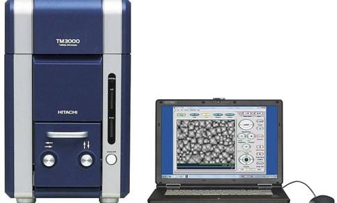
A scanning electron microscope (SEM) uses a high-energy beam of electrons to scan a specimen rather than using optical (visible) light (electromagnetic radiation). An electron gun accelerates electrons toward an anode which collimates the electrons into a beam of parallel “rays” of electrons. A circular magnet then focuses the electron beam on the specimen. The detectors capture the backscattered electrons, and a computer program electronically assimilates the detections into an image. All of this must happen in a vacuum or else the electrons are scattered by air molecules producing random “noise”. We cannot use wet samples because SEM uses a vacuum, and it will varporize water. Also, we cannot use live specimens because they will die either due to the vacuum or due to the bombardment of electrons. If samples are to be preserved, they should be plated with a very thin layer of gold. The gold plating also produces a better resolution and depth of field, but this process is expensive.